Stimulated emission depletion (STED) microscopy
It is a process that provides super resolution by selectively deactivating fluorophores, so as to enhance the imaging in that area. It was developed by Stefan W. Hell in 1994, and was first experimentally shown in 1999. Hell was awarded the Nobel Prize in Chemistry in 2014 for its invention. This is one of several types of super resolution microscopy techniques that have recently been developed. Super resolution microscopy is a set of techniques to bypass the diffraction limit of microscopy to achieve better resolution.
Photoactivated localization microscopy (PALM) and stochastic optical reconstruction microscopy (STORM) are also super resolution microscopy techniques, although they use a different process than STED to achieve this resolution.

The techniques they developed enabled extremely high resolution images to be produced using optical microscopy. Their work circumvented the problem of the ‘diffraction limit’ – the inability of light microscopy to distinguish between structures smaller than half the wavelength of visible light or about 200nm. This advance allowed nanoscale structures – including individual molecules – to be visualised within cells while they are still alive, something that isn’t possible with techniques such as electron microscopy.
The basis of STED microscopy is the coupling of the excitation laser with the STED depletion laser, resulting in the doughnut-shaped depletion. The two perfectly aligned laser systems minimize the size of the fluorescence spot, overcoming the resolution-limiting effects of diffraction.
The perhaps most straightforward way to sharpen the fluorescence focal spot is to selectively inhibit the fluorescence at its outer part. If this is applied to an otherwise diffraction-limited spot, one would expect that the diffraction barrier can be overcome since scanning with a smaller fluorescent spot signifies increased spatial resolution. A phenomenon that stops fluorescence (=spontaneous emission) is that of stimulated emission. This is one of the key ingredients of the Stimulated Emission Depletion (STED-) microscope. However, STED by itself could not really break the diffraction barrier since the beams with which STED is accomplished are diffraction-limited as well. Therefore the real physical ingredient for breaking the diffraction barrier is the saturation of the fluorescence inhibition by stimulated emission, as we will argue below.
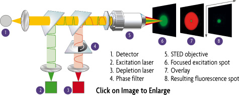
 The setup of STED Microscope
The STED-microscope relies on pairs of synchronized laser pulses. To this end, excitation is performed by a subpicosecond laser pulse that is tuned to the absorption spectrum of the dye. The excitation pulse is focused into the sample, producing an ordinary diffraction limited spot of excited molecules. The excitation pulse is immediately followed by a depletion pulse, dubbed 'STED-pulse'. The STED pulse is red-shifted in frequency to the emission spectrum of the dye, so that its lower energy photons act ideally only on the excited dye molecules, quenching them to the ground state by stimulated emission. The net effect of the STED pulse is that the affected excited molecules cannot fluoresce because their energy is dumped and lost in the STED pulse. By spatially arranging the STED pulse in a doughnut mode, only the molecules at the periphery of the spot are ideally quenched . In the center of the doughnut, where the STED pulse is vanishing, fluorescence ideally remains unaffected.
No resolution limit For Microscopy
By increasing the STED pulse intensity, the depletion becomes complete at the spot's periphery and increasingly more effective towards the middle. At the doughnut hole, however, the fluorescence is ideally not affected at all. Therefore, by increasing the intensity of the doughnut-shaped STED-pulse, the fluorescent spot can be progressively narrowed down, in theory, even to the size of a molecule. This concept signifies a fundamental breaking of the diffraction barrier. The essential ingredient is the saturated reduction of the fluorescence (= depletion) at any coordinate but the focal point.
 Comparison with confocal fluorescence microscopy
This microscopy is in stark contrast to the presently known super resolution methods like the confocal, the multiphoton or related fluorescence microscopes, which can never surpass Abbe's barrier by more than a factor 2. In a way, confocal fluorescence and two-photon microscopes just cross the diffraction border, without breaking it. The resolution of these systems is still limited by diffraction, in contrast to the STED-microscope .
Depletion means saturation
The real physical reason for the breaking of the diffraction barrier is not the fact that fluorescence is inhibited, but the saturation (of the fluorescence reduction). Fluorescence reduction alone would not help since the focused STED-pulse is also diffraction-limited. What does saturation mean in this context? Whereas the fluorescence at the middle of the doughnut is unaffected, it is fully stopped at the closest proximity of the doughnut. Thus the fluorescent region is continuously narrowed down without limit!
Fundamentally enlarged passband of the optical transfer function
It is clear that the decrease in spatial extent of the effective spot or point-spread-function in a STED-microscope is associated with a fundamental increase of the passband of the effective transfer function of the microscope. The STED-microscope is not a diffraction-limited system anymore. It is the first to provide conceptionally unlimited optical resolution, in spite of the fact that it relies on visible light and regular objective lenses .

To date an improvement beyond the diffraction barrier of 3 in the transverse direction and up to 6 along the optical axis has been experimentally demonstrated. The viability of the STED-concept has been exemplified in a number of simple experiments. Its practicability and the maximum spatial resolution depend very much on the level of saturation that can be obtained and on the deepness of the doughnut hole, which should be ideally zero. So far, experiments show that the level of saturation will be determined by the bleaching that is inflicted on the dye. Moreover, it will be interesting to see to which extent dyes can be switched off and if STED is applicable to all dyes, including those that are endogenous to the cell.
Ground-State-Depletion- (GSD) Microscopy, a cousin of STED
An alternative to quenching the excited state is to deplete the ground state of the dye. This depletion could be achieved by shelving the dye into the triplet state or another long-lived state. As in the concept of STED, the real ingredient is the saturation of the depletion. Saturation entails a non-linear relationship between the (residual) fluorescence and the applied intensity
|


